DEPARTMENT OF CHEMISTRY AND BIOCHEMISTRY OF NUCLEOPROTEINS

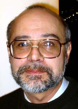
Andrey B. Vartapetian
Head of Department, Sc.D., Professor
Tel.: +7(495)939-4125
E-mail: varta@belozersky.msu.ru
Department of Chemistry and Biochemistry of Nucleoproteins was founded in 1969 by Academician of RAS Prof. Alexey A. Bogdanov. Since 2003 the Department is headed by Prof. Andrey B. Vartapetian..
The research focus of the Department teams is in the field of molecular and cellular biology. Investigations are aimed at the elucidation of molecular mechanisms of protein biosynthesis, programmed cell death, and cell proliferation.
The Department includes:
The main areas of research in the Department are:
Laboratory of Protein Synthesis Regulation
Staff: Ivan N. Shatsky - Head of Laboratory, Professor, D. Sci.; Sergey E. Dmitriev - Researcher, Ph.D.; Ilya M. Terenin - Junior Researcher, Ph.D.; Dmitriy E. Andreev - Junior Researcher, Ph.D.
The laboratory is concerned with investigations of molecular mechanisms of translation initiation and its regulation in mammalian cells. The main approach developed and used by the laboratory is the in vitro reconstitution of translation initiation complexes from totally purified components (1). Using this method, the mechanisms of internal translation initiation on the IRES-elements of some picornavirus and hepatitis C virus RNAs have been elucidated (1, 2). In collaborative researches with the lab of Dr. M. Hentze (EMBL, Heidelberg, Germany), the mechanism of lipoxygenase mRNA silencing in erythroid differentiation has been determined (3). An important role of translation initiation factor eIF4B in the differential expression of mammalian mRNAs has been proposed (4). During the last years, a unique mechanism of translation initiation on the cross-kingdom IRES-element of Rhopalosiphum padi virus RNA has been deciphered (5). It has been found for the first time that so-called leaderless mRNAs (i.e. mRNAs lacking 5'UTRs) are capable of programming 80S ribosomes in the absence of translation initiation factors (6). At present, the lab is focused on the mechanism of translation initiation of the natural dicistronic mRNA transcribed from human retrotransposon LINE and on the nature of the interaction of hepatitis C virus IRES with mammalian 40S ribosomal subunits (7).
Selected Recent Publications::
1. Pestova, T.V., Hellen, C.U.T., and Shatsky, I.N. 1996. Canonical eukaryotic
initiation factors determine initiation of translation by internal ribosomal
entry. Mol.Cell. Biol. 16, 6859-6869.
2. Pestova, T.V., Shatsky, I.N., Fletcher, S.P., Jackson, R.J., and Hellen,
C.U.T. 1998. A prokaryotic-like mode of cytoplasmic eukaryotic ribosome
binding to the initiation codon during internal translation initiation of
hepatitis C and classical swine fever virus RNAs. Genes Dev. 12, 67-83.
3. Ostareck, D.H., Ostareck-Lederer, A., Shatsky, I.N., and Hentze, M.
2001. Lipoxygenase mRNA silencing in erythroid differentiation:The 3'UTR
regulatory complex controls 60S ribosomal subunit joining. Cell 104, 281-290.
4. Dmitriev, S.E., Terenin, Y.M., Dunaevsky, Y.E., Merrick, W.C., and
Shatsky, I.N. 2003. Assembly of 48S translation initiation complexes from
purified components with mRNAs that have some base-pairing within their 5’-UTRs.
Mol. Cell. Biol. 23, 8925-8933.
5. Terenin, I.M., Dmitriev, S.E., Andreev, D.E., Royall, E., Belsham,
G.J., Roberts, L.O., and Shatsky, I.N. 2005. A “cross-kingdom” IRES reveals
a simplified mode of internal ribosome entry. Mol. Cell. Biol. 25, 7879-7888.
6. Andreev, D.E., Terenin, I.M., Dunaevsky, Y.E., Dmitriev, S.E., and
Shatsky, I.N. 2006. A leaderless mRNA can bind to mammalian 80S ribosomes
and direct polypeptide synthesis in the absence of translation initiation
factors. Mol. Cell. Biol. 26, 3164-3169.
7. Laletina, E., Graifer, D., Malygin, A., Ivanov, A., Shatsky, I., and
Karpova, G. 2006. Proteins surrounding hairpin IIIe of the hepatitis C virus
internal ribosome entry site on the human 40S ribosomal subunit. Nucleic
Acids Res. 34, 2027-2036.
Laboratory of Molecular Biology of the Gene
Staff: Andrey B. Vartapetian - Head of the Laboratory, Professor, D.Sci.; Alexandra G. Evstafieva - Leading Researcher, Ph.D.; Nina V. Chichkova - Senior Researcher, Ph.D.; Raisa A. Galiullina - Researcher, Ph.D.; Grigoriy S. Filonov - Researcher; Sergey V. Melnikov - Graduate Student; Tatyana N. Chernysheva - techn.; Elena S. Yukhina - techn.
The main directions of research in the laboratoryare:
Animal Direction:
A group headed by Dr. A. Evstafieva studies structure-function relationships in human nuclear protein ProT?. Apart from its proliferation-related function, ProT? was shown to be involved in protection of human cells from oxidative stress. ProT? acts as an intranuclear 'dissociator' of the complex formed by the key transcription factor Nrf2 with its inhibitor protein Keap1. By specific binding to Keap1, ProT? liberates Nrf2 to activate oxidative stress-protecting genes. In accord with this model, Keap1 was shown to be a nuclear-cytoplasmic shuttling protein equipped with a potent nuclear export signal. Furthermore, Keap1 shuttling governs intracellular distribution of Nrf2.
In collaboration with the laboratory of Prof. V. Agol (Belozersky Institute), apoptosis-related properties of ProT? were identified. ProT? was shown to defend human cells from apoptosis. However when protection is surmounted, ProT? is quickly truncated by caspase-3 and losses its C-terminal region encompassing the nuclear localization signal. As a consequence, truncated ProT? re-localizes from the nucleus to the cytoplasm and further to the surface of the dying cell, thus becoming a specific surface marker of apoptotic cells.
Plant Direction:
A group headed by Dr. N. Chichkova has identified a functional analogue of animal caspases in plants. In collaboration with the laboratory of Dr. M.Taliansky (SCRI, Dundee, UK) it was shown that, although the plant enzyme is structurally different from animal caspases, it possesses all of the properties of animal caspases. The plant enzyme is a highly specific proteinase with cleavage specificity similar to that of human caspase-3. The enzyme is dormant in healthy plant cells and becomes activated upon the induction of programmed cell death in plants. Activity of this enzyme is essential for the accomplishment of plant PCD. Several protein targets of plant caspase have been identified.
Selected Recent Publications::
1. Sukhacheva E.A., Evstafieva A.G., Fateeva T.V., Shakulov V.R., Efimova N.A., Karapetian R.N., Rubtsov Y.P., and Vartapetian A.B. (2002) Sensing prothymosin alpha origin, mutations and conformation with monoclonal antibodies. J. Immunol. Methods 266, 185-196.
2. Evstafieva A.G., Belov G.A., Rubtsov Y.P., Kalkum M., Joseph B., Chichkova N.V., Sukhacheva E.A., Bogdanov A.A., Pettersson R.F., Agol V.I., and Vartapetian A.B. (2003) Apoptosis-related fragmentation, translocation, and properties of human prothymosin alpha. Exp. Cell Res. 284, 209-221.
3. Vartapetian A.B. and Uversky V.N. (2003) Prothymosin alpha: a simple yet mysterious protein. In: Protein Structures: Kaleidoscope of Structural Properties and Functions (V.N.Uversky, ed.) Research Signpost, Kerala, pp. 223-237.
4. Yang C.H., Murti A., Baker S., Frangou-Lazaridis M., Vartapetian A.B., Murti K.G., and Pfeffer L.M. (2004) Interferon induces the interaction of prothymosin ? with STAT3 and results in the nuclear translocation of the complex. Exp. Cell Res. 298, 197-206.
5. Chichkova N.V., Kim S.H., Titova E.S., Kalkum M., Morozov V.S., Rubtsov Y.P., Kalinina N.O., Taliansky M.E., and Vartapetian A.B. (2004) A plant caspase-like protease activated during the hypersensitive response. Plant Cell 16, 157-171.
6. Karapetian R.N., Evstafieva A.G., Abaeva I.S., Chichkova N.V., Filonov G.S., Rubtsov Y.P., Sukhacheva E.A., Melnikov S.V., Schneider U., Wanker E.E., and Vartapetian A.B. (2005) Nuclear oncoprotein prothymosin ? is a partner of Keap1: Implications for expression of oxidative stress-protecting genes. Mol. Cell. Biol. 25, 1089-1099.
7. Reavy B., Bagirova S., Chichkova N.V., Fedoseeva S.V., Kim S.H., Vartapetian A.B., and Taliansky M.E. (2007) Caspase-resistant VirD2 protein provides enhanced gene delivery and expression in plants. Plant Cell Rep. 26, 1215-1219.
8. Reavy B., Taliansky M., Kim S.H., Vartapetian A.B., Chichkova N.V., Bagirova S. (2007) Modified VirD2 protein and its use in improved gene transfer. WO/2007/132193.
9. Chichkova N.V., Galiullina R.A., Taliansky M.E., Vartapetian A.B. (2008) Tissue disruption activates a plant caspase-like protease with TATD cleavage specificity. Plant Stress 2, 89-95.
Laboratory of Nucleic Acid-Protein Interactions
Staff: Yuri F. Drygin - Head of Laboratory, D.Sci.; Elena S. Gavrushina - techn.
The laboratory studies a natural covalent complex between the VPg protein and genomic RNA of encephalomyocarditis virus.
Recent Publications:
1. Yusupova RA, Gulevich AY, Drygin YF. 2000. Isolation from ascites carcinoma Krebs II cells of an unlinking enzyme hydrolyzing a covalent bond between picornavirus RNA and VPg. Biochemistry (Mosc). 65:1219-26.
2. Gulevich AY, Yusupova RA, Drygin YF. 2001. A phosphodiesterase from ascites carcinoma Krebs II cells specifically cleaves the bond between VPg and RNA of encephalomyocarditis virus. Biochemistry (Mosc). 66:345-9.
3. Gulevich AY, Yusupova RA, Drygin YF. 2002. VPg unlinkase, the phosphodiesterase that hydrolyzes the bond between VPg and picornavirus RNA: a minimal nucleic moiety of the substrate. Biochemistry (Mosc). 67:615-21.
4. Ivanova OA, Venyaminova AG, Repkova MN, Drygin YF. 2005. Polyclonal antibodies against a structure mimicking the covalent linkage unit between picornavirus RNA and VPg: an immunochemical study. Biochemistry (Mosc). 70:1038-45.
Research Group of Structure-Function Analysis of Ribosomes
Staff: Alexey A. Bogdanov - Group Leader, Academician of RAS, Professor; Yuri S. Sharanov - Junior Researcher.
The group investigates mechanisms of functional signal transduction in the elongation cycle of the ribosome (in collaboration with the Chair of Chemistry of Natural Compounds, Chemical Department, Moscow State University).
Selected Recent Publications:
1. Sergiev PV, Bogdanov AA, Dahlberg AE, Dontsova O. 2000. Mutations at position A960 of E. coli 23 S ribosomal RNA influence the structure of 5 S ribosomal RNA and the peptidyltransferase region of 23 S ribosomal RNA. J Mol Biol. 299, 379-389.
2. Matadeen R, Sergiev P, Leonov A, Pape T, van der Sluis E, Mueller F, Osswald M, von Knoblauch K, Brimacombe R, Bogdanov A, van Heel M, Dontsova O. 2001. Direct localization by cryo-electron microscopy of secondary structural elements in Escherichia coli 23 S rRNA which differ from the corresponding regions in Haloarcula marismortui. J Mol Biol. 307, 1341-1349.
3. Zvereva MI, Ivanov PV, Teraoka Y, Topilina NI, Dontsova OA, Bogdanov AA, Kalkum M, Nierhaus KH, Shpanchenko OV. 2001. Complex of transfer-messenger RNA and elongation factor Tu. Unexpected modes of interaction. J Biol Chem. 276, 47702-47708.
4. Leonov AA, Sergiev PV, Bogdanov AA, Brimacombe R, Dontsova OA. 2003. Affinity purification of ribosomes with a lethal G2655C mutation in 23 S rRNA that affects the translocation. J Biol Chem. 278, 25664-25670.
5. Shpanchenko OV, Zvereva MI, Ivanov PV, Bugaeva EY, Rozov AS, Bogdanov AA, Kalkum M, Isaksson LA, Nierhaus KH, Dontsova OA. 2005. Stepping transfer messenger RNA through the ribosome. J Biol Chem. 280, 18368-18374.
6. Sergiev PV, Lesnyak DV, Burakovsky DE, Kiparisov SV, Leonov AA, Bogdanov AA, Brimacombe R, Dontsova OA. 2005. Alteration in location of a conserved GTPase-associated center of the ribosome induced by mutagenesis influences the structure of peptidyltransferase center and activity of elongation factor G. J Biol Chem. 280, 31882-31889.
7: Sergiev PV, Lesnyak DV, Kiparisov SV, Burakovsky DE, Leonov AA, Bogdanov AA, Brimacombe R, Dontsova OA. 2005. Function of the ribosomal E-site: a mutagenesis study. Nucleic Acids Res. 33, 6048-6056.
Animal Cell Culture Group
Staff: Alexander N. Kuimov - Group Leader, Ph.D.; Natalya N. Sidorova - techn., M.S.
The subject of our research is an enzyme tankyrase (a human isoenzyme tankyrase 2). The enzyme catalyzes posttranslational protein poly(ADP-ribosylation), yet its physiological role is not clear. We have identified tankyrase 2 as a tumor antigen. Furthermore, we have shown for the first time that tankyrase 2 is also localized in some normal human tissues like the epithelium of renal tubules and intestine. We suggest that the enzyme participates in food uptake in intestine and reabsorption process in renal tubules. We use methods of biochemistry and enzymology, and the source of our enzyme is a culture of epithelial human kidney cells. This is why we support the cell culture service in our institute and help in promotion of the cell culture methods in scientific practice. Read more about Animal Cell Culture Laboratory at:
http://www.genebee.msu.su/anb/accl/EACCLab/homepage.htm
Recent Publications:
1. Boitchenko VE, Korobko VG, Prassolov VS, Kravchenko VV, Kuimov AN, Turetskaya RL, Kuprash DV, Nedospasov SA. 2000. Immunodetection of Murine Lymphotoxins in Eukaryotic Cells. Russ J Immunol. 5:259-266.
2. Kuimov AN, Kuprash DV, Petrov VN, Vdovichenko KK, Scanlan MJ, Jongeneel CV, Lagarkova MA, Nedospasov SA. 2001. Cloning and characterization of TNKL, a member of tankyrase gene family. Genes Immun. 2:52-5.
3. Kuimov AN, Terekhov SM. 2003. Soluble tankyrase located in cytosol of human embryonic kidney cell line 293. Biochemistry (Mosc). 68:260-8.
4: Kuimov AN. 2004. Polypeptide components of telomere nucleoprotein complex. Biochemistry (Mosc). 69:117-29.
5. Sidorova N, Zavalishina L, Kurchashova S, Korsakova N, Nazhimov V, Frank G, Kuimov A. 2006. Immunohistochemical detection of tankyrase 2 in human breast tumors and normal renal tissue. Cell Tissue Res. 323:137-45.